The Human Anatomy and Physiology
THE HUMAN ORGANISM
Chemical Level
click image to enlarge
The structural and functional characteristics of all organisms are determined by their cellular makeup.their chemical level of organization involves interaction between atoms,which are tiny building blocks of matter.Atoms can combine to form molecules,such as water,sugar,fats,proteins,and deoxyribose nucleic acid (DNA).The function of a molecule is related intimately to its structure.For example,collagen molecules are strong,ropelike fibers that give skin structural strength and flexibility.With old age,the structure of collagen changes,and the skin becomes fragile and torn more easily.
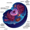
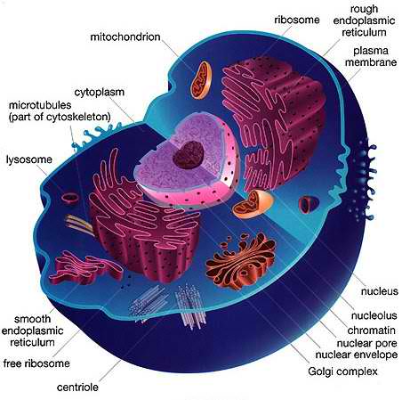
Cellular Level
the animal cell(celula animals)
click image to enlarge
click image to enlarge
Cells are basic structural and functional units of organisms,such as plants and animals.Molecules can combine to form organelles,which are the small structures that make up cells.for example,the nucleus contains the cell's hereditary information and mitochondria manufacture adenosine triphosphate (ATP),which is used by cells as a source of energy.although cell types differ in their structure and funtion,they have many characteristics in common.knowledge of these characteristics and their variations is essential to basic understanding of anatomy and physiology.
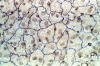
Tissue Level
simple squamous epithelial tissue
click image to enlarge

click image to enlarge
A tissue is a group of similar cells and the materials surrounding them.The characteristics of the cells and surrounding materials determinehte functions of the tissue.The many tissues that make up the body are classified into four tissue types:epithelial,connective,muscle and nervous.
Organ Level
left kidney
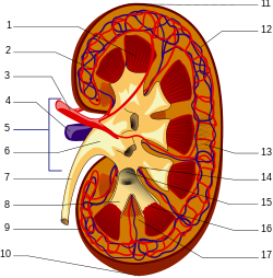
An organ is composed of two or more tissue types that together perform one or more common functions.the urinar bladder,skin,stomach,eye,heart are examples of organs.
Organ System Level
human skeletal system
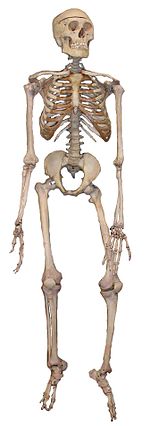
An organ system is a group of organs classified as a unit because of a common function or set of functions.For example,the urinary system consist of the kidneys,ureter,urinary bladder, and urethra.The kidney produces urine,which is transported by the ereters to the urinary bladder,where it is stored until eliminated from the body by passing through the urethra.
Organism Level

An organism is any livng thing considered as a whole-whether composed of one cell,such as bacterium,or trillions if cells,such as humans.The human organnism is a complex of organ systems that are mutually dependent on one another.
HOMEOSTASIS
=>Is the maintanance of a relatively constant environment within the body.
=>Is the maintanance of a relatively constant environment within the body.
SET POINT
=>homeostatic mechanisms normally maintain the normal value.
That normal value is called "Set Point".
=>homeostatic mechanisms normally maintain the normal value.
That normal value is called "Set Point".
NORMAL RANGE
=>note that these mechanisms are not able to maintain precisely at the set point.
instead,a range that is when mechanisms remain within these range,hemeostasis is maintained.
=>note that these mechanisms are not able to maintain precisely at the set point.
instead,a range that is when mechanisms remain within these range,hemeostasis is maintained.
That range is the NORMAL RANGE.
NEGATIVE FEEDBACK
=>Any deviation from the set point is made smaller or resisted.
negative feedback does not prevent deviation within the normal range.
NEGATIVE FEEDBACK
=>Any deviation from the set point is made smaller or resisted.
negative feedback does not prevent deviation within the normal range.
Example : Maintanance of blood pressure. normal blood pressure is necesssary for the movement of heart to the tissues.
the blood supplies the tissues with oxygen and nutrients and removes waste products,thus maintaining tissue homeostasis.
the blood supplies the tissues with oxygen and nutrients and removes waste products,thus maintaining tissue homeostasis.
POSITIVE FEEDBACK
=>When a deviation from a normal values occurs,the response to the system is to make the deviation even greater.
Example:inadequate delivery of blood to cardiac.contraction of cardiac muscle generartes blood pressure and moves blood
through blood vessels to tissues.A system of blood vessels on the outside of the heart produces cardiac muscle with blood supply
sufficient to allow normaal contranctions to occur.
but with extreme blood loss, inadequation to blood results the heart pumps less blood,causing death to organs and cells.
DIRECTIONAL TERMS
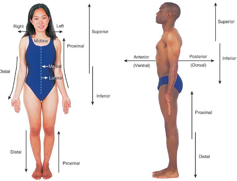
SUPERIOR-INFERIOR
those words denote vertical levels of position
>a structure located above is superior.
example:the mouth is superior to chin.
>a structure located below is inferior.
example:chin is inferior to mouth.
ANTERIOR-POSTERIOR
>Anterior is the one that is the most forward to surface of the body.
example:canine teeth are anterior to premolars.
>posterior is the one that is following the anterior,much like its opposite.
example:premolars are posterior to canine teeth(notice that it was much like just vice versa?)
CEPHALIC-CAUDAL
>cephalic is an anatomical direction that is close to head
>caudal is an antomical direction that pertains to posterior end of body
DORSAL-VENTRAL
>Dorsal is a direction to the back(back of head,back of chest,etc.)
>ventral is a direction to the front(chest,palms,etc.)
PROXIMAL-DISTAL
>Point of reference of attachment to other part of body
(proximal-one nearer to attachment)
(distal-one farther to attachment)
MEDIAL-LATERAL
>Medial is the one closest to the midline of the body.
(midline is the imaginative vertical line at the center of the body.)
>lateral is the one farther to the midline of the body.
BODY PLANES

SAGITTAL SECTION
divides the body into left and right portion by cutting
the midsagittal part(a cut in midline.)
TRANSVERSE SECTION
(It is also known as a cross section)
cuts the body into top and bottom part by cutting
the body in horizontal cut.
FRONTAL SECTION
divides the body into front and back part.
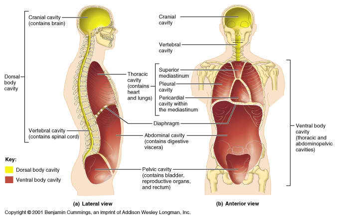
BODY CAVITIES
CELL STRUCTURE AND THEIR FUNCTIONS
CELL STRUCTURE AND THEIR FUNCTIONS
Plasma membrane
Outer boundary of cells that controls
entry and exit of substances
attaches to other cells or intercellular
molecules;part of intercellular communication
and identification;catalyzes chemical reactions
Cytoplasm
contains enzymes that catalyze the synthesis
and breakdown of molecules.
Cytoskeletons
support the cytoplasm and form centrioles,
spindle fibers cilia and flagella
provide structural support to cells.
Nuclear envelope
seperates nucleus from cytoplasm;allows movement
of materials into and out of nucleus.
Chromatin
DNA regulates protein synthesis and the
chem. reactions of the cell;DNA is the genetic or
hereditary material.
Nucleolus
Assembly site of large and small ribosomal units.
Ribosome
Site of protein synthesis
Rough Endoplasmic Reticulum
protein synthesis and transport to golgi apparatus.
Smooth Endoplasmic Reticulum
manufactures lipids and carbohydrates;detoxifies
harmful chemicals;store calcium.
Golgi Apparatus
modifies proteins and lipids and packages them
into vescicles for distribution
Secretory Vesicle
carries proteins to cell surface for secretion.
Lysosomes
Contains digestive enzymes
for food vacuoles "suicide sac".
Mitochondria
Major site of ATP synthesis.
Centrioles
Centers for microtubule formation;determine
cell polarity during cell division.
Spindle fibers
Assists in the separation of chromosomes during cell
division.
Cilia
Move materials over the surface of cells.
Flagellum
Responsible for movement of sperm cells.
Microvilli
Increase surface surface area of the plasma membrane
for absorption and secretion
Two Major Kinds of Cell
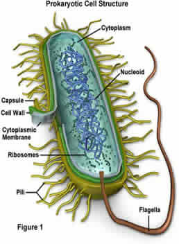
Prokaryotic Cell
Cells that do not have a cell membrane around their Nucleus. Example - Bacteria

Eukaryotic Cell
Cells that have a membrane around their nucleus. Example - Plant and Animal Cells
Type Of Movement Across The Cell Membrane
Passive Transport
Passive transport is the movement of molecules
across the cell membrane and does not require energy.
It is dependent on the permeability of the cell membrane.
There are three main kinds of passive transport -
Diffusion, Osmosis and Facilitated Diffusion.

Diffusion
The movement of molecules from a region of higher
concentration to a region of lower concentration.
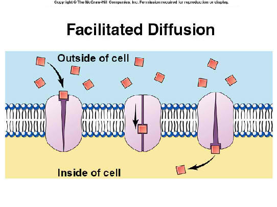
Facilitated diffusion
This process does not require ATP but does require cell membrane
proteins which are called carrier proteins to carry the molecules
across the cell membrane from an area of higher concentration
to an area of lower concentration.

Osmosis
The movement of water across a semi permeable membrane.
Osmosis is the movement of water (red dots) through a
semipermeable membrane to a higher concentration of
solutes (blue dots).
click image to enlarge
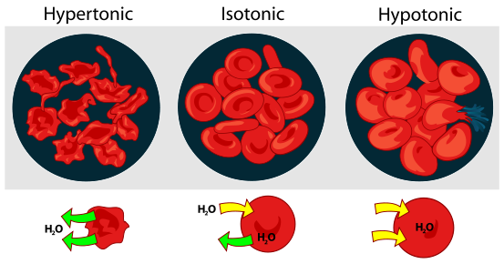
Hypertonic Solution
A Hypertonic solution contain a high concentration
of solute in relation to the solution within the cell
(e.g. the cell's cytoplasm).
When a cell is placed in a hypertonic solution, the water
diffuses out of the cell, causing the cell to shrivel up.
Hypotonic Solution
A hypotonic solution contain A solution
with a lower salt concentration than in normal cells
When a cell is placed in a hypotonic solution,
the water diffuses into the cell, causing the
cell to swell and possibly explode.
Isotonic Solution
A solution that has the same salt concentration
as the normal cells of the body and the blood.
When a cell is placed in an isotonic solution,
the water diffuses into and out of the cell at the same
rate. The fluid that surrounds the body cells is isotonic.
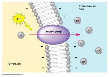
Active Transport
Active Transport requires the cell to use energy,
usually in the form of ATP.
Active Transport creates a charge gradient
in the cell membrane. For example in the mitochondrion,
hydrogen ion pumps pump hydrogen ions into the intermembrane
space of the organelle as part of making ATP.
Active Transport keeps unwanted ions or other molecules out of the
cell that are able to diffuse through the cell membrane.
Active transport uses energy to send substances against
the direction they would travel by simple diffusion:
that is from a region of low concentration to a region
of high concentration.
Moving other Materials and Substances
Into and out of the cell.
ENDOCYTOSIS and EXOCYTOSIS
ENDOCYTOSIS
Endocytosis (Endo (within) cytosis (cell) )
is a process in which a substance (e.g. proteins)
gains entry into a cell without passing through
the cell membrane.
EXOCYTOSIS
Endocytosis (Exo (exit) cytosis (cell) ) is a
process in which a substance is exited from the cell
without passing through the cell membrane.
Examples of thigs that migh be exited include
secretion of proteins like enzymes, hormones
and antibodies.
Phases Of Mitosis

click image to enlarge
1. Interphase
DNA has replicated, but has not formed
the condensed structure of chromosome.
They remain as loosely coiled chromatin.
The nuclear membrane is still intact
to protect the DNA molecules from undergoing mutation.
2. Prophase
The DNA molecules progressively shorten and
condense by coiling, to form chromosomes.
The nuclear membrane and nucleolus are no longer visible.
The spindle apparatus has migrate to opposite poles of the cell..
3. Metaphase
The spindle fibres attach themselves to the centromeres of
the chromosomes and align the the chromosomes at the equatorial plate.
4. Anaphase
The spindle fibres shorten and the centromere splits,
separated sister chromatids are pulled along behind the centromeres.
5.Telophase
The chromosomes reach the poles of their respective spindles.
Nuclear envelope reform before the chromosomes uncoil.
The spindle fibres disintegrate.
Protein Synthesis,Transcription and Translation
Steps in Protein Synthesis:
STEP 1: The first step in protein synthesis is the transcription of mRNA
from a DNA gene in the nucleus. At some other prior time, the various
other types of RNA have been synthesized using the appropriate DNA.
The RNAs migrate from the nucleus into the cytoplasm.
STEP 2: Initiation:
In the cytoplasm, protein synthesis is actually initiated by the AUG
codon on mRNA. The AUG codon signals both the interaction of the ribosome
with m-RNA and also the tRNA with the anticodons (UAC). The tRNA which
initiates the protein synthesis has N-formyl-methionine attached.
The formyl group is really formic acid converted to an amide
using the -NH2 group on methionine (left most graphic)
The next step is for a second tRNA to approach the mRNA (codon - CCG).
This is the code for proline. The anticodon of the proline tRNA
which reads this is GGC. The final process is to start growing peptide
chain by having amine of proline to bond to the carboxyl acid group of
methinone (met) in order to elongate the peptide.
STEP 3: Elongation:
Elongation of the peptide begins as various tRNA's read the next codon.
In the example on the left the next tRNA to read the mRNA is tyrosine.
When the correct match with the anticodons of a tRNA has been found,
the tyrosine forms a peptide bond with the growing peptide chain .
The proline is now hydrolyzed from the tRNA. The proline tRNA now moves
away from the ribosome and back into the cytoplasm to reattach another
proline amino acid.
Step 4: Elongation and Termination:
When the stop signal on mRNA is reached, the protein synthesis is terminated.
The last amino acid is hydrolyzed from its t-RNA.
The peptide chain leaves the ribosome. The N-formyl-methionine that was used
to initiate the protein synthesis is also hydrolyzed from the completed peptide
at this time.
The ribosome is now ready to repeat the synthesis several more times.
TISSUES, GLANDS, AND MEMBRANES
Functions Of Epithelia
Protection: Convering epithelium specially
of skin oral cavity and esphagus protect
underline structures.
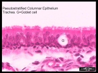
Secretion: Goblet cells and Gastric mucosal
surface and othres do the function of secretion.
Absorption: In kidney and small intestine
selective things are absorbed.
Filtration: Urine, sweat and carbon dioxide
are examples.
Excretion: Certain epithelial cells do this from
the blood those waste products which are carried
in the blood. Urine and sweat and carbon dioxide
are examples of this also.
Classification Of Epithelium
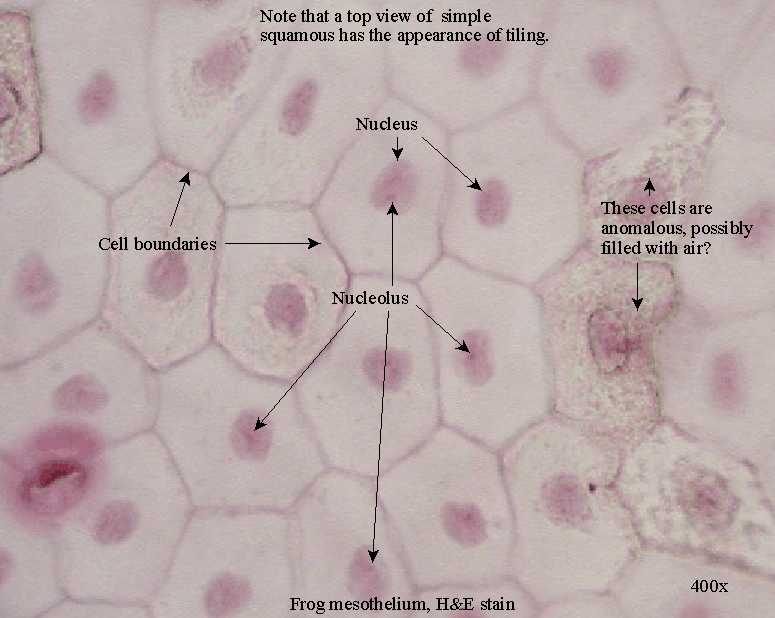
Simple Squamous Epithelium
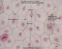
Function: Diffusion,Filtration,Secretion and
Some protection against friction.
Location:Alveoli of lungs,linig of blood
vessels,inner surface of the eardrum
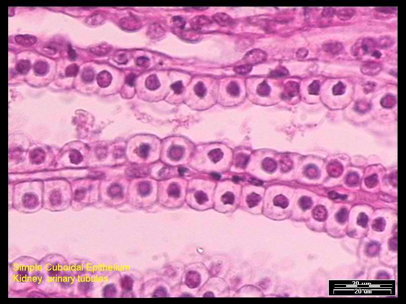
Simple Cuboidal Epithelium
Functions:active transport and facilitated
diffusion result in secretion and absorption
by cells of the kidney tubules.
Location:kidney tubules,glands and their ducts,
choroid plexuses of the brain,surface of the ovaries
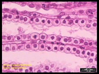
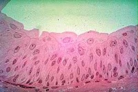
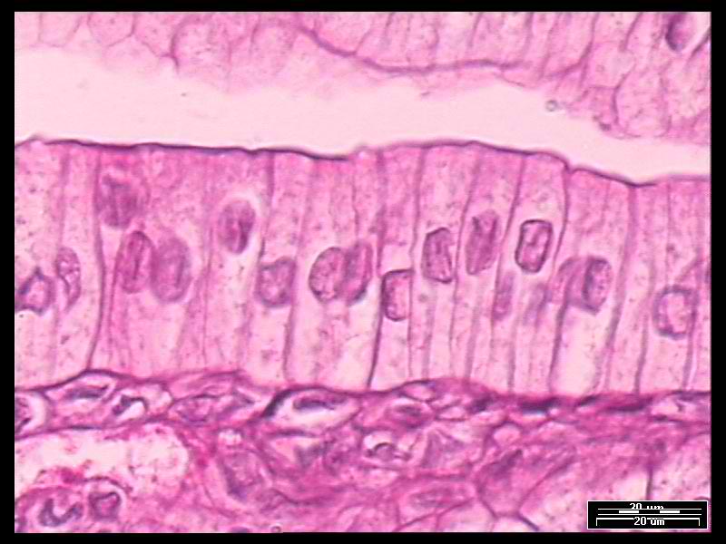
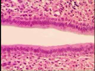
Simple Columnar Epithelium
Function:movement,absorption,
secretion
Location:brochioles of lungs,
uterus,uterine tubes

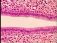
Pseudostratified Columnar Epithelium
Function:secretion,filtration
Location:lining of nasal cavity,bronchi
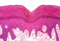
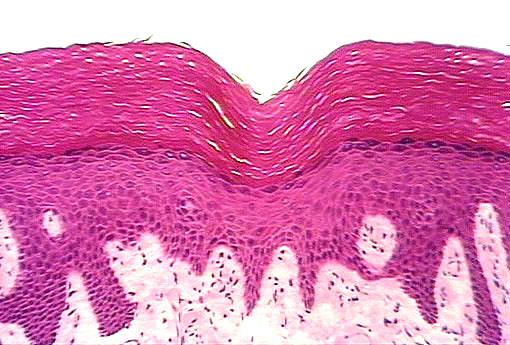
Stratified Squamous Epithelium
Functions:protection,barrier,reducer
Location:keratinized-skin,non-keratinized-mouth,
throat,larynx
Stratified Cuboidal Epithelium
Function:secretion,absorption,protection
Location:sweat gland,salivary gland

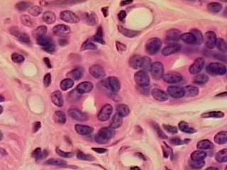
Stratified Columnar
Function:protection and secretion
Location:mammary gland duct,male urethra
Transitional Epithelium
Function:protection,fluctuation
Location:bladder,ureter,urethra
In epithelium or tissues,there is a secretory
structure that is called "glands".
glands are responsible for secretion of anti bodies,
mucous,and dead cells.
Protection: Convering epithelium specially
of skin oral cavity and esphagus protect
underline structures.
Secretion: Goblet cells and Gastric mucosal
surface and othres do the function of secretion.
Absorption: In kidney and small intestine
selective things are absorbed.
Filtration: Urine, sweat and carbon dioxide
are examples.
Excretion: Certain epithelial cells do this from
the blood those waste products which are carried
in the blood. Urine and sweat and carbon dioxide
are examples of this also.
Classification Of Epithelium
Simple Squamous Epithelium

Function: Diffusion,Filtration,Secretion and
Some protection against friction.
Location:Alveoli of lungs,linig of blood
vessels,inner surface of the eardrum
Simple Cuboidal Epithelium
Functions:active transport and facilitated
diffusion result in secretion and absorption
by cells of the kidney tubules.
Location:kidney tubules,glands and their ducts,
choroid plexuses of the brain,surface of the ovaries
Simple Columnar Epithelium
Function:movement,absorption,
secretion
Location:brochioles of lungs,
uterus,uterine tubes
Pseudostratified Columnar Epithelium
Function:secretion,filtration
Location:lining of nasal cavity,bronchi

Stratified Squamous Epithelium
Functions:protection,barrier,reducer
Location:keratinized-skin,non-keratinized-mouth,
throat,larynx
Stratified Cuboidal Epithelium
Function:secretion,absorption,protection
Location:sweat gland,salivary gland
Stratified Columnar
Function:protection and secretion
Location:mammary gland duct,male urethra
Transitional Epithelium
Function:protection,fluctuation
Location:bladder,ureter,urethra

In epithelium or tissues,there is a secretory
structure that is called "glands".
glands are responsible for secretion of anti bodies,
mucous,and dead cells.
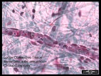
Connective tissues
Loose Connective Tissue
Function:loose packing,
support,nourishment
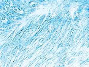
Dense regular collageneous tissue
Function:withstand pressure and forces
Location:tendon and ligament

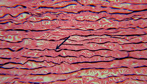
Dense Regular Elastic Tissue
Function:stretching and recoiling
Location:Vocal Folds
Dense Irregular Connective Tissue
Function:tensile strength capable in
stretching in all directions
Location:dermis of skin
Dense Irregular Elastic
Function:capable of stretching and recoil
in several directions
Location:elastic arteries
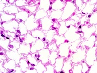
Adipose Tissue
Function:packing material,
thermal insulator,energy storage
protection
Location:mammary glands
bone marrow
Reticular Tissue
Function:superstructure
for lymph
Location:lymph nodes

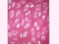
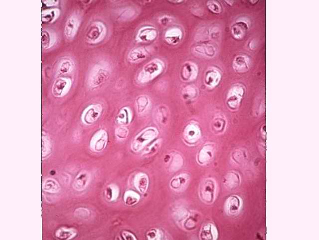
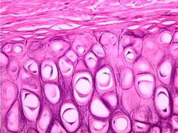
Hyaline Cartilage
Function:growth,rigidity and
flexibility
Location:long bones,costal cartilage
Fibrocartilage
Function:flexible and
can withstand pressure
Location:intervertebral disks

Elastic Cartilage
Function:provides rigidity with
more flexibility
Location:ears,auditory tubes

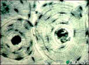
Bone
Function:Provides great strength and support
Location:Bones

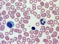
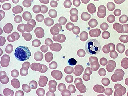
Blood
Function:trnsports oxygen,carbon dioxide
hormones nutrients
Location:blood vessels and heart
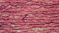
Muscle Tissues
Skeletal muscle
Function:movement of body
Location:attached to bone
![Skeletal muscle 03a[1]](http://theultimateanatomy.xtgem.com/images/Skeletal%20muscle%2003a[1]_thumb.jpg)
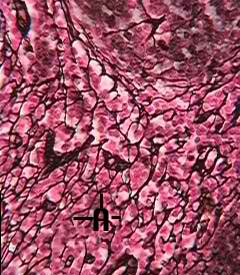
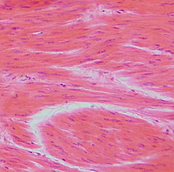
Cardiac Muscle
Function:pumps blood
Location:heart
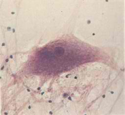
Smooth Muscle
Functions:regulates size nad contraction
Location:lining of stomach
Neurons Or Neuroglia
Functions:trnasmit information
Location:brain,spinal cord,ganglia
Loose Connective Tissue
Function:loose packing,
support,nourishment

Dense regular collageneous tissue
Function:withstand pressure and forces
Location:tendon and ligament
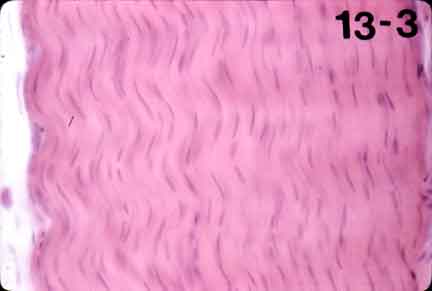
Dense Regular Elastic Tissue
Function:stretching and recoiling
Location:Vocal Folds

Dense Irregular Connective Tissue
Function:tensile strength capable in
stretching in all directions
Location:dermis of skin
Dense Irregular Elastic
Function:capable of stretching and recoil
in several directions
Location:elastic arteries
Adipose Tissue
Function:packing material,
thermal insulator,energy storage
protection
Location:mammary glands
bone marrow

Reticular Tissue
Function:superstructure
for lymph
Location:lymph nodes
Hyaline Cartilage
Function:growth,rigidity and
flexibility
Location:long bones,costal cartilage
Fibrocartilage
Function:flexible and
can withstand pressure
Location:intervertebral disks
Elastic Cartilage
Function:provides rigidity with
more flexibility
Location:ears,auditory tubes
Bone
Function:Provides great strength and support
Location:Bones
Blood
Function:trnsports oxygen,carbon dioxide
hormones nutrients
Location:blood vessels and heart
Muscle Tissues
Skeletal muscle
Function:movement of body
Location:attached to bone
![Skeletal muscle 03a[1]](http://theultimateanatomy.xtgem.com/images/Skeletal%20muscle%2003a[1].jpg)
Cardiac Muscle
Function:pumps blood
Location:heart
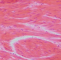
Smooth Muscle
Functions:regulates size nad contraction
Location:lining of stomach
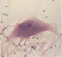
Neurons Or Neuroglia
Functions:trnasmit information
Location:brain,spinal cord,ganglia
THE INTEGUMENTARY SYSTEM
The Integumentary system has many functions:
Protects the body's internal living tissues
and organs
Protects against invasion by infectious
organisms
Protects the body from dehydration
Protects the body against abrupt changes
in temperature
Helps dispose of waste materials
Acts as a receptor for touch, pressure,
pain, heat and cold
Stores water, fat, and vitamin D.
Parts Of The Skin
The Epidermis
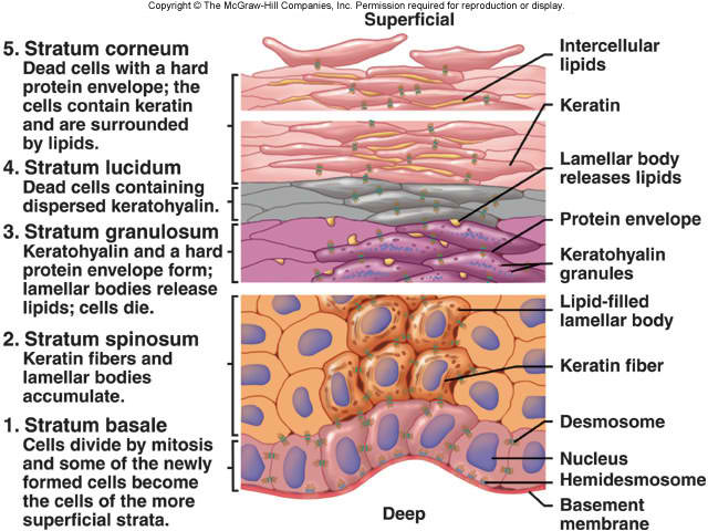
STRATUM BASALE
The stratum basale (basal layer, sometimes referred to as stratum germinativum) is the deepest layer of the five layers of the epidermis, which is the outer covering of skin in mammals. The stratum basale is a continuous layer of cells. It is often described as one cell thick, though it may in fact be two to three cells thick in glabrous (hairless) skin and hyperproliferative epidermis (from a skin disease).
The stratum basale is primarily made up of basal keratinocyte cells, which can be considered the stem cells of the epidermis. They divide to form the keratinocytes of the stratum spinosum, which migrate superficially.
STRATUM SPINOSUM
The stratum spinosum (or spinous layer) is a
layer of the epidermis found between the stratum granulosum and stratum basale. This layer is also referred to as the "spinous" or "prickle-cell" layer. This appearance is due to desmosomal connections of adjacent cells. Keratinization begins in the stratum spinosum.
STRATUM GRANULOSUM
The stratum granulosum (or granular layer) is a thin layer of cells in the epidermis.Keratinocytes migrating from the underlying stratum spinosum become known as granular cells in this layer. These cells contain keratohyalin granules, protein structures that promote hydration and crosslinking of keratin.
At the transition between this layer and the stratum corneum, cells secrete lamellar bodies (containing lipids and proteins) into the extracellular space. This results in the formation of the hydrophobic lipid envelope responsible for the skin's barrier properties
STRATUM LUCIDUM
The stratum lucidum (Latin for "clear layer") is a thin, clear layer of dead skin cells in the epidermis named for its translucent appearance under a microscope. It is found only in areas of thick skin, most noticeably on the palms of the hands and the soles of the feet.
Located between the stratum granulosum and stratum corneum layers, it is composed of three to five layers of dead, flattened keratinocytes. The keratinocytes of the stratum lucidum do not feature distinct boundaries and are filled with eleidin, an intermediate form of keratin.
The thickness of the lucidum is controlled by the rate of mitosis of the epidermal cells. In addition, melanocytes determine the darkness of the stratum lucidum. The cells of the stratum lucidum are flattened and contain an oily substance that is the result of exocytosis of lamellar bodies accumulated while the keratinocytes are moving through the stratum spinosum and stratum granulosum.
STRATUM CORNEUM
The stratum corneum (Latin for horned layer) is the outermost layer of the epidermis, consisting of dead cells (corneocytes) that lack nuclei and organelles. The purpose of the stratum corneum is to form a barrier to protect underlying tissue from infection, dehydration, chemicals and mechanical stress. Desquamation, the process of cell shedding from the surface of the stratum corneum, balances proliferating keratinocytes that form in the stratum basale. These cells migrate through the epidermis towards the surface in a journey that takes approximately fourteen days.
Skin Color
Melanocytes are melanin-producing cells located in the bottom layer (the stratum basale) of the skin's epidermis, the middle layer of the eye (the uvea), the inner ear, meninges,bones, and heart. Melanin is a pigment that is responsible primarily for the color of skin.
Through a process called melanogenesis, these cells produce melanin, which is a pigment found in the skin, eyes, and hair. This melanogenesis leads to a long-lasting pigmentation, which is in contrast to the pigmentation that originates from oxidation of already-existing melanin.
There are both basal and activated levels of melanogenesis; in general, lighter-skinned people have low basal levels of melanogenesis. Exposure to UV-B radiation causes an increased melanogenesis as a response to DNA photodamage.
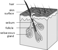
The hair
Papilla
At the base of the follicle is a large structure that is called the papilla.
The papilla is made up mainly of connective tissue and a capillary loop. Cell division in the papilla is either rare or non-existent.
Matrix
Around the papilla is the hair matrix, a collection of epithelial cells often interspersed with the pigment-producing cells, melanocytes. Cell division in the hair matrix produces the cells that will form the major structures of the hair fiber and the inner root sheath. The hair matrix epithelium is one of the fastest growing cell populations in the human body, which is why some forms of chemotherapy that kill dividing cells or radiotherapy may lead to temporary hair loss. The papilla is usually ovoid or pear shaped with the matrix wrapped completely around it except for a short stalk-like connection to the surrounding connective tissue that provides access for the capillary.
Root sheath
The root sheath is composed of an external and internal root sheath. The external root sheath appears empty with cuboid cells when stained with H&E stain. The internal root sheath is composed of three layers, Henle's layer, Huxley's layer, and an internal cuticle that is continuous with the outermost layer of the hair fiber.
Hair fiber
The hair fiber is composed of keratin.
Bulge
The bulge is located in the outer root sheath at the insertion point of the arrector pili muscle. It houses several types of stem cells, which supply the entire hair follicle with new cells, and take part in healing the epidermis after a wound.
Other structures
Other structures associated with the hair follicle include arrector pili muscles, sebaceous glands, and apocrine sweat glands. Hair follicle receptors sense the position of the hairs.
Attached to the follicle is a tiny bundle of muscle fiber called the arrector pili. This muscle is responsible for causing the follicle lissis to become more perpendicular to the surface of the skin, and causing the follicle to protrude slightly above the surrounding skin (piloerection) and a pore encased with skin oil. This process results in goose bumps (or goose flesh).
Also attached to the follicle is a sebaceous gland, which produces the oily or waxy substance sebum. The thicker the density of the hair, the more sebaceous glands that are found.
NAIL
A nail is a horn-like envelope covering the
dorsal aspect of the terminal phalanges of
fingers and toes in humans
The matrix (synonyms: matrix unguis, keratogenous membrane, nail matrix, onychostroma) is the tissue (or germinal matrix) upon which the nail rests, the part of the nail bed that extends beneath the nail root and contains nerves, lymph and blood vessels. The matrix is responsible for the production of the cells that become the nail plate. The width and thickness of the nail plate is determined by the size, length, and thickness of the matrix, while the shape of the fingertip itself determines if the nail plate is flat, arched or hooked. The matrix will continue to grow as long as it receives nutrition and remains in a healthy condition. As new nail plate cells are incubated, they emerge from the matrix round and white to push older nail plate cells forward; and in this way yet older cells become compressed, flat, and translucent, making the pink colour of the capillaries in the nail bed below visible.
The lunula (occasionally called simply "the moon") is the visible part of the matrix, the whitish crescent-shaped base of the visible nail. The lunula is largest in the thumb and often absent in the little finger.
The nail bed is the skin beneath the nail plate.Like all skin, it is composed of two types of tissues: the deeper dermis, the living tissue fixed to the bone which contains capillaries and glands, and the superficial epidermis, the layer just beneath the nail plate which moves forward with the plate. The epidermis is attached to the dermis by tiny longitudinal "grooves" known as the matrix crests or crests of nail matrix (cristae matricis unguis). During old age, the plate thins and these grooves are made evident in the structure.
The nail sinus (sinus unguis) is the deep furrow into which the nail root is inserted.[
The nail root (radix unguis) is the part of nail situated in the nail sinus, i.e. the base of the nail embedded underneath the skin. It originates from the actively growing tissue below, the matrix.
The nail plate or body of nail (corpus unguis) is the actual nail, and like hair and skin, made of translucent keratin protein made of amino acids. In the nail it forms a strong flexible material made of several layers of dead, flattened cells. The plate appears pink because of the underlying capillaries. Its (transversal) shape is determined by the form of the underlying bone. In common usage, the word nail often refers to this part only.
The free margin (margo liber) or distal edge is the anterior margin of the nail plate corresponding to the abrasive or cutting edge of the nail. The hyponychium (informally known as the "quick") is the epithelium located beneath the nail plate at the junction between the free edge and the skin of the fingertip. It forms a seal that protects the nail bed. The onychodermal band is the seal between the nail plate and the hyponychium. It is found just under the free edge, in that portion of the nail where the nail bed ends and can be recognized by its glassy, greyish colour (in fair-skinned people). It is not perceptible in some individuals while it is highly prominent on others.
The eponychium is the small band of epithelium that extends from the posterior nail wall onto the base of the nail. Often and erroneously called the "proximal fold" or "cuticle", the eponychium is the end of the proximal fold that folds back upon itself to shed an epidermal layer of skin onto the newly formed nail plate. This layer of non-living, almost invisible skin is the cuticle that "rides out" on the surface of the nail plate. Together, the eponychium and the cuticle form a protective seal. The cuticle on the nail plate is dead cells and is often removed during manicure, but the eponychium is living cells and should not be touched. The perionyx is the projecting edge of the eponychium covering the proximal strip of the lunula.
The nail wall (vallum unguis) is the cutaneous fold overlapping the sides and proximal end of the nail. The lateral margin (margo lateralis) is lying beneath the nail wall on the sides of the nail and the nail groove or fold (sulcus matricis unguis) are the cutaneous slits into which the lateral margins are embedded.
The paronychium is the border tissue around the nail and paronychia is an infection in this area.
Protects the body's internal living tissues
and organs
Protects against invasion by infectious
organisms
Protects the body from dehydration
Protects the body against abrupt changes
in temperature
Helps dispose of waste materials
Acts as a receptor for touch, pressure,
pain, heat and cold
Stores water, fat, and vitamin D.
Parts Of The Skin
The Epidermis
STRATUM BASALE
The stratum basale (basal layer, sometimes referred to as stratum germinativum) is the deepest layer of the five layers of the epidermis, which is the outer covering of skin in mammals. The stratum basale is a continuous layer of cells. It is often described as one cell thick, though it may in fact be two to three cells thick in glabrous (hairless) skin and hyperproliferative epidermis (from a skin disease).
The stratum basale is primarily made up of basal keratinocyte cells, which can be considered the stem cells of the epidermis. They divide to form the keratinocytes of the stratum spinosum, which migrate superficially.
STRATUM SPINOSUM
The stratum spinosum (or spinous layer) is a
layer of the epidermis found between the stratum granulosum and stratum basale. This layer is also referred to as the "spinous" or "prickle-cell" layer. This appearance is due to desmosomal connections of adjacent cells. Keratinization begins in the stratum spinosum.
STRATUM GRANULOSUM
The stratum granulosum (or granular layer) is a thin layer of cells in the epidermis.Keratinocytes migrating from the underlying stratum spinosum become known as granular cells in this layer. These cells contain keratohyalin granules, protein structures that promote hydration and crosslinking of keratin.
At the transition between this layer and the stratum corneum, cells secrete lamellar bodies (containing lipids and proteins) into the extracellular space. This results in the formation of the hydrophobic lipid envelope responsible for the skin's barrier properties
STRATUM LUCIDUM
The stratum lucidum (Latin for "clear layer") is a thin, clear layer of dead skin cells in the epidermis named for its translucent appearance under a microscope. It is found only in areas of thick skin, most noticeably on the palms of the hands and the soles of the feet.
Located between the stratum granulosum and stratum corneum layers, it is composed of three to five layers of dead, flattened keratinocytes. The keratinocytes of the stratum lucidum do not feature distinct boundaries and are filled with eleidin, an intermediate form of keratin.
The thickness of the lucidum is controlled by the rate of mitosis of the epidermal cells. In addition, melanocytes determine the darkness of the stratum lucidum. The cells of the stratum lucidum are flattened and contain an oily substance that is the result of exocytosis of lamellar bodies accumulated while the keratinocytes are moving through the stratum spinosum and stratum granulosum.
STRATUM CORNEUM
The stratum corneum (Latin for horned layer) is the outermost layer of the epidermis, consisting of dead cells (corneocytes) that lack nuclei and organelles. The purpose of the stratum corneum is to form a barrier to protect underlying tissue from infection, dehydration, chemicals and mechanical stress. Desquamation, the process of cell shedding from the surface of the stratum corneum, balances proliferating keratinocytes that form in the stratum basale. These cells migrate through the epidermis towards the surface in a journey that takes approximately fourteen days.
STRATUM CORNEUM
The stratum corneum (Latin for horned layer) is the outermost layer of the epidermis, consisting of dead cells (corneocytes) that lack nuclei and organelles. The purpose of the stratum corneum is to form a barrier to protect underlying tissue from infection, dehydration, chemicals and mechanical stress. Desquamation, the process of cell shedding from the surface of the stratum corneum, balances proliferating keratinocytes that form in the stratum basale. These cells migrate through the epidermis towards the surface in a journey that takes approximately fourteen days.
Skin Color
Melanocytes are melanin-producing cells located in the bottom layer (the stratum basale) of the skin's epidermis, the middle layer of the eye (the uvea), the inner ear, meninges,bones, and heart. Melanin is a pigment that is responsible primarily for the color of skin.
Through a process called melanogenesis, these cells produce melanin, which is a pigment found in the skin, eyes, and hair. This melanogenesis leads to a long-lasting pigmentation, which is in contrast to the pigmentation that originates from oxidation of already-existing melanin.
There are both basal and activated levels of melanogenesis; in general, lighter-skinned people have low basal levels of melanogenesis. Exposure to UV-B radiation causes an increased melanogenesis as a response to DNA photodamage.
The hair
Papilla
At the base of the follicle is a large structure that is called the papilla.
The papilla is made up mainly of connective tissue and a capillary loop. Cell division in the papilla is either rare or non-existent.
Matrix
Around the papilla is the hair matrix, a collection of epithelial cells often interspersed with the pigment-producing cells, melanocytes. Cell division in the hair matrix produces the cells that will form the major structures of the hair fiber and the inner root sheath. The hair matrix epithelium is one of the fastest growing cell populations in the human body, which is why some forms of chemotherapy that kill dividing cells or radiotherapy may lead to temporary hair loss. The papilla is usually ovoid or pear shaped with the matrix wrapped completely around it except for a short stalk-like connection to the surrounding connective tissue that provides access for the capillary.
Root sheath
The root sheath is composed of an external and internal root sheath. The external root sheath appears empty with cuboid cells when stained with H&E stain. The internal root sheath is composed of three layers, Henle's layer, Huxley's layer, and an internal cuticle that is continuous with the outermost layer of the hair fiber.
Hair fiber
The hair fiber is composed of keratin.
Bulge
The bulge is located in the outer root sheath at the insertion point of the arrector pili muscle. It houses several types of stem cells, which supply the entire hair follicle with new cells, and take part in healing the epidermis after a wound.
Other structures
Other structures associated with the hair follicle include arrector pili muscles, sebaceous glands, and apocrine sweat glands. Hair follicle receptors sense the position of the hairs.
Attached to the follicle is a tiny bundle of muscle fiber called the arrector pili. This muscle is responsible for causing the follicle lissis to become more perpendicular to the surface of the skin, and causing the follicle to protrude slightly above the surrounding skin (piloerection) and a pore encased with skin oil. This process results in goose bumps (or goose flesh).
Also attached to the follicle is a sebaceous gland, which produces the oily or waxy substance sebum. The thicker the density of the hair, the more sebaceous glands that are found.

NAIL
A nail is a horn-like envelope covering the
dorsal aspect of the terminal phalanges of
fingers and toes in humans
The matrix (synonyms: matrix unguis, keratogenous membrane, nail matrix, onychostroma) is the tissue (or germinal matrix) upon which the nail rests, the part of the nail bed that extends beneath the nail root and contains nerves, lymph and blood vessels. The matrix is responsible for the production of the cells that become the nail plate. The width and thickness of the nail plate is determined by the size, length, and thickness of the matrix, while the shape of the fingertip itself determines if the nail plate is flat, arched or hooked. The matrix will continue to grow as long as it receives nutrition and remains in a healthy condition. As new nail plate cells are incubated, they emerge from the matrix round and white to push older nail plate cells forward; and in this way yet older cells become compressed, flat, and translucent, making the pink colour of the capillaries in the nail bed below visible.
The lunula (occasionally called simply "the moon") is the visible part of the matrix, the whitish crescent-shaped base of the visible nail. The lunula is largest in the thumb and often absent in the little finger.
The nail bed is the skin beneath the nail plate.Like all skin, it is composed of two types of tissues: the deeper dermis, the living tissue fixed to the bone which contains capillaries and glands, and the superficial epidermis, the layer just beneath the nail plate which moves forward with the plate. The epidermis is attached to the dermis by tiny longitudinal "grooves" known as the matrix crests or crests of nail matrix (cristae matricis unguis). During old age, the plate thins and these grooves are made evident in the structure.
The nail sinus (sinus unguis) is the deep furrow into which the nail root is inserted.[
The nail root (radix unguis) is the part of nail situated in the nail sinus, i.e. the base of the nail embedded underneath the skin. It originates from the actively growing tissue below, the matrix.
The nail plate or body of nail (corpus unguis) is the actual nail, and like hair and skin, made of translucent keratin protein made of amino acids. In the nail it forms a strong flexible material made of several layers of dead, flattened cells. The plate appears pink because of the underlying capillaries. Its (transversal) shape is determined by the form of the underlying bone. In common usage, the word nail often refers to this part only.
The free margin (margo liber) or distal edge is the anterior margin of the nail plate corresponding to the abrasive or cutting edge of the nail. The hyponychium (informally known as the "quick") is the epithelium located beneath the nail plate at the junction between the free edge and the skin of the fingertip. It forms a seal that protects the nail bed. The onychodermal band is the seal between the nail plate and the hyponychium. It is found just under the free edge, in that portion of the nail where the nail bed ends and can be recognized by its glassy, greyish colour (in fair-skinned people). It is not perceptible in some individuals while it is highly prominent on others.
The eponychium is the small band of epithelium that extends from the posterior nail wall onto the base of the nail. Often and erroneously called the "proximal fold" or "cuticle", the eponychium is the end of the proximal fold that folds back upon itself to shed an epidermal layer of skin onto the newly formed nail plate. This layer of non-living, almost invisible skin is the cuticle that "rides out" on the surface of the nail plate. Together, the eponychium and the cuticle form a protective seal. The cuticle on the nail plate is dead cells and is often removed during manicure, but the eponychium is living cells and should not be touched. The perionyx is the projecting edge of the eponychium covering the proximal strip of the lunula.
The nail wall (vallum unguis) is the cutaneous fold overlapping the sides and proximal end of the nail. The lateral margin (margo lateralis) is lying beneath the nail wall on the sides of the nail and the nail groove or fold (sulcus matricis unguis) are the cutaneous slits into which the lateral margins are embedded.
The paronychium is the border tissue around the nail and paronychia is an infection in this area.